How should healthy iPSC colonies look? What characteristics define a bad colony?
Example of a crater formation in hiPSC colonies, not healthy!

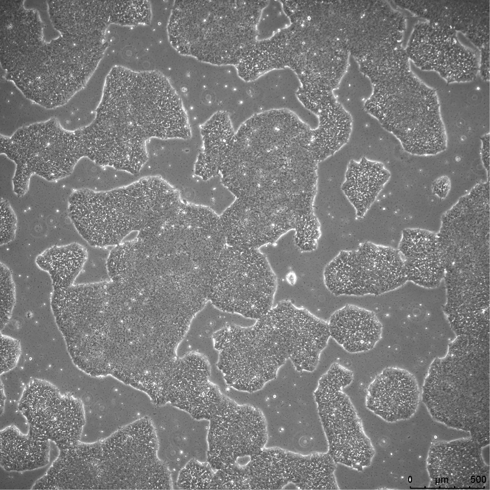
Healthier hiPSC colonies below, notice the smooth edges:
hiPSC colonies undergoing differentiation, look for triangular or spiky cells at edges:

Underconfluence of hiPSC colonies:

Is there somewhere on the AICS website where I can find a collection of healthy and unhealthy colony images suitable for benchmarking a machine-learning based classifier?
Hi @gregj, We do not have a set at this moment available on our website. However, maybe one of our scientists may be able to provide images. About how many images of healthy and unhealthy would you need for machine learning?
Thanks!
Hello,
I have recently thawed an Allen Cell line and saw the “crater-like” morphology in the first image. Do you have any insights into what causes this morphology? And how to minimize its occurrence?
Thank you!
Hello,
There are several reasons I can think of that could explain your less than ideal morphology. It could be your lot of Matrigel and/or mTESR supplement is the issue. We actually lot test both of these to ensure our cell morphology stays consistent and purchase large quantities to keep things as uniform as we can. It could also be your cell morphology will improve as they recover from the thaw and do a recovery passage or two. Sometimes you might see mild cratering until the colonies become more mature after a split but by day 3 or 4, they will look well packed and healthy. If they are still in Rock inhibitor, also give it a day out of it to reassess their morphology. Hopefully, this helps and your cells recover well!